
Page 6 - Scialabba_alii_1999
P. 6
•
9
,o . 116
.;o 9 7 04
o''" 66
~o· 45
30 •
oo .. /'; 27 • 29
-: ·: " ~·:: 22 ~
: 21 )
..
cc
,o
- ,. 1 1 1403
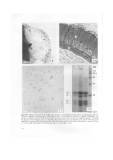
Figg. 9/12 o9) Nearly ripe seed. Pheno l droplets in the radiai and external peripheral walls (arrows) of mucilaginous cells (mc).
Column (c). 10) Ripe ~eed imegumems with the following layers: a) mucilaginous: b) crushed subepidermal: c) palisade; d) pigo
memed; e) aleurone: f) hyaline. Il ) Ripe seed. Transverse ;ection of embryonic axis showing phosphatase activity in intercellu-
lar spaces (arrows) of conica! cylinder (cc). 12) SDS polyacrylamide electrophoretic separation from seeds al different stages of
maturatio n: (a) green seed; (b) hard green-brown seed; (c) ripe brown sced; (d) molecular weight ;tandard proteins in KD. The
arrows on the left indicate proteins with major quantitativc variations. Ban,: IO !111l.
152