
Page 3 - vol64-2011-215-222deidun
P. 3
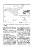
Figure 1. Distribution of the sampled localities. The codes of the sampled sites correspond to those reported in table 1.
  Triangles denote beaches where P. bimaculata was collected, whilst squares denote beaches where P. acuminata
  was collected.

placed in each trap as an attractant, with the traps being  viduals, fifteen of which were collected from the
deployed at dusk and emptied at dawn. Collected Phale-      beaches of San Blas and Xatt l-Ahmar in Gozo (Maltese
ria spp. specimens were sorted out and fixed in situ in     Islands - MAL), fifteen from Gelso beach on the island
80% ethanol.                                                of Vulcano (Aeolian Islands - VUL), and fifteen from
                                                            Oliveri beach (SIC), along the northern coast of Sicily.
  Identification of the collected specimens was carried     The head and forelegs were dissected from the speci-
out under a stereomicroscope according to the morpho-       mens to be used for future molecular analyses. After
logical characteristics listed in Canzoneri (1968). When    dissection, the remaining body parts were mounted on a
necessary, selected specimens were dissected in order to    card.
study the form of the aedeagus.
                                                              The specimens were positioned along a horizontal
Geometric morphometrics                                     plane; for each individual, the right portion of the body
  Geometric morphometrics techniques, nowadays a            was examined. Dorsal imagines were digitized using a
                                                            Leica D-LUX 3<<LMS>> camera mounted on the opti-
standard protocol in morphological research and de-         cal stereomicroscope Wild M3.
scribed in Rohlf and Marcus, 1993; Adams et al., 2004
and Zelditch et al., 2004, were used to analyse the intra-    The digital imagines of the pronotum and the elytra
specific differentiation in the shape of pronotum and the   were processed separately with MakeFan6 software
elytra in three populations of P. bimaculata. We focused    (Sheets, 2003). Within the pronotum, the cartesian x, y
on these two body parts because the shape of pronotum       coordinates of four landmarks and six semi-landmarks
and elytra is considered of diagnostic interest in the ge-  were recorded. In the elytra, the cartesian x, y coordi-
nus Phaleria. Moreover, previous studies in tenebrionid     nates of four landmarks and twelve semi-landmarks
beetles showed that pronotum shape is particularly suit-    were recorded. The position of the pronotum landmarks
able for geometric morphometric analyses (Palmer,           and semi-landmarks adopted in this study is shown in
2002a; 2002b; Taravati et al., 2009). Statistical varia-    figure 2a, while the position of elytra landmarks and
tions in the shape of the pronotum and the right elytra in  semi-landmarks is shown in figure 2b.
different P. bimaculata individuals from different popu-
lations were analyzed using multivariate statistics.          The bi-dimensional coordinates of the anatomical
                                                            landmarks and semi-landmarks on the outline of the
  Analyses were performed on 45 P. bimaculata indi-         dorsal view of the pronotum and the right elytra were

                                                            217