
Page 5 - Coletti_al2016
P. 5
Carnets Geol. 16 (3)
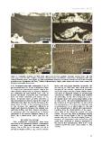
Figure 2: Vegetative anatomy. (A) Thick crust, upper part of Lower Langhian, Romania, Lopadea Veche. (B) Thin
crust, upper part of Lower Langhian, Romania, Lopadea Veche. (C) Plumose ventral core, Burdigalian, NW Italy,
Tertiary Piedmont Basin, Torre Veglio. (D) High-magnification detail of a secondary ventral-core and of the overlying
peripheral zone, Burdigalian, NW Italy, Tertiary Piedmont Basin, Uviglie; white arrow=cell fusion; black arrows=cell
filaments with visible primary pits.
2.A). Peripheral zone cells rectangular, 8 to 31 tacles with very large diameter (generally 700
μm in length and 8 to 15 μm in diameter (Table μm but up to 1000 μm) may result from the
2). Primary pit connections connect cells of the merging of two nearby reproductive chambers
same filament while cells of adjacent filaments since such large structures were always obser-
are connected by cells fusions (Fig. 2.D); in ved close to adjacent couples of normal-sized
Recent specimens secondary pits connections conceptacles with the separating wall partially
have not been observed (see BASSO et al., dissolved (Fig. 3.E-F). The conceptacles protru-
2011). Trichocytes have not been observed. It de slightly above the surrounding thallus surfa-
was not possible to observe clearly the shape of ce (Fig. 3.A-F). The H/D (height/diameter) ran-
epithallial cells either due to the abrasion of the ges between 0.25 and 0.5 (Table 2). The roof of
upper layer of cells or the excessive thickness the conceptacle is 27 to 62 μm in thickness and
of the sections. No significant difference in is composed of 3 to 6 cells (Table 2). The roof
length was observed between the alleged sub- of the conceptacles is pitted with depressions,
epithallial initials and their inward derivatives, resulting from the disintegration of the upper-
in agreement with the observations of Recent most cells of the filaments around the pore
specimens of the species (NÓBREGA-FARIAS et al., canals. The roof pits have a diameter ranging
2010, Fig. 4; BASSO et al., 2011, Figs. 6 & 10- from 19 to 59 μm (Table 2; Fig. 3). The dege-
11). nerate cells are 9 to 38 μm in length (Table 2).
Rosettes of degenerate cells around the pore
REPRODUCTIVE ANATOMY canal were observed in sections tangential to
the roof of the conceptacles, with 6 to 7 cells
Only multiporate conceptacles have been counted around the pore canal (Table 2; Fig.
observed in the specimens studied. Conceptacle 3.G-H).
chambers are generally sub-rectangular in
shape, 260 to 550 μm in diameter and 90 to
190 μm in height (Table 2; Fig. 3.A-D). Concep-
31