
Page 3 - Brain_morphology_2008
P. 3
Original Paper
Series of four sections were cut every 50 µm of tis- a
sue; in each series, one of the sections was stained
with one of the four antisera. For each primary anti-
serum, the percentage of immunoreactive cells was
estimated by counting the cells with a clearly visible
nucleus in 18 immunostained sections.
The count was performed using light microscopy
fields captured at 20x magnification and digitalized
with a Quantimet 500W image analyser. Data are
given as means ± standard deviation.
b
Results
Encephalization quotient and gross brain mor-
phology
The mean brain mass (EA) of the bluefin tuna
sampled was 6.52±1.78 g and the mean body mass
was 97.1±19.8 kg. Based on the brain mass to body Figure 1. Photographs of bluefin tuna brain. a) dorso-lateral
mass relationship obtained for several fish species aspect; b) ventral aspect. Bar = 2 cm.
by Lisney and Collin (2006), the expected brain
mass (EE) of the bluefin tuna specimens sampled in
the present investigation was calculated to be 9.25
g.Therefore, the QE is <1 i.e. 6.52/9.25 = 0.71.
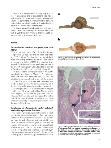
The dorsal-lateral and ventral views of the bluefin
tuna brain are shown in Figure 1. The olfactory
bulbs are not well developed and, in fact, are
attached to the rostral-ventral part of the olfacto-
ry lobes. The optic tecta are well developed and
exhibit a deep groove. The corpus cerebelli, which
displays a remarkable rostral-caudal extension dor-
sal to the optic tecta up to the medulla oblongata,
exhibits an evident external folding. The octavolat-
eralis regions, the eminentia granularis and the
crista cerebelli are also well developed.The ventral
view of the brain is characterized by a very large
hypophysis caudally followed by pre-eminent hypo-
thalamic inferior lobes.
Morphology of diencephalic nuclei putatively
involved in reproductive functions
The nucleus preopticus-periventricularis is local-
ized in the diencephalon starting from its rostral
end where it covers the inner surface of the ependy-
ma lining the wall of the relevant ventricle (III ven-
tricle or median cavity) (Figure 2). This nucleus
consists of a few sparse neurons in the rostral part,
whereas its aboral part is constituted by numerous Figure 2. Micrographs of bluefin tuna brain showing the nucle-
us periventricularis, the nucleus preopticus and the cavity of
cells closely packed and arranged in several layers the third ventricle (bar = 500 µm). Inset: higher magnification
(the nucleus periventricularis itself) (Figure 2).The of the nucleus periventricularis (bar = 100 µm). Asterisk, cavi-
ty of the third ventricle; npo, nucleus preopticus; npv, nucleus
ventral part of the nucleus periventricularis bends periventricularis. Haematoxylin-Eosin staining.
21