
Page 5 - Brain_morphology_2008
P. 5
Original Paper
distinct neuron populations in terms of cell size: i)
small cells with a minor axis of 10.5±2.5 µm and
a major axis of 18.0±4.1 µm; ii) large cells with a
minor axis of 22.3±4.5 µm and a major axix of
32.7±6.9 µm.These neurons, usually arranged in 3-
6 layers, are interspersed in a close net of blood
capillaries (Figure 2) and exhibit a characteristic
topographic distribution within the nucleus. In the
vertical part of the nucleus (nucleus periventricu-
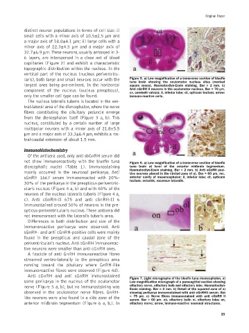
laris), both large and small neurons occur with the Figure 5. a) Low magnification of a transverse section of bluefin
tuna brain showing the oculomotor nucleus sites (marked
largest ones being pre-eminent. In the horizontal square zones). Haematoxilyn-Eosin staining. Bar = 2 mm. b)
component of the nucleus (nucleus preopticus), Anti cGnRH II neurons in the oculomotor nucleus. Bar = 70 µm.
cv, cerebelli valvula; il, inferior lobe; ot, opticum tectum; arrow:
only the smaller cell type can be found. immuno-reactive cells.
The nucleus lateralis tuberis is located in the ven-
tral-lateral area of the diencephalon,where the nerve
fibres constituting the pituitary peduncle emerge
from the diencephalon itself (Figure 3 a, b). This
nucleus, constituted by a certain number of large
multipolar neurons with a minor axis of 21.8±5.5
µm and a major axis of 33.3±6.4 µm, exhibits a ros-
tral-caudal extension of about 1.5 mm.
Immunohistochemistry
Of the antisera used, only anti sbGnRH serum did
not show immunoreactivity with the bluefin tuna Figure 6. a) Low magnification of a transverse section of bluefin
diencephalic nuclei (Table 1). Immunostaining tuna brain at level of the anterior midbrain tegmentum.
Haematoxilyn-Eosin staining. Bar = 2 mm. b) Anti sGnRH posi-
mainly occurred in the neuronal perikarya. Anti tive neurons placed in the circled zone of a). Bar = 60 µm. mc,
sGnRH 1667 serum immunoreacted with 20%- anterior cavity of mesencephalon; il, inferior lobe; ot, opticum
tectum; asterisk, recessus lateralis.
30% of the perikarya in the preopticus-periventric-
ularis nucleus (Figure 4 a, b) and with 40% of the
neurons of the nucleus lateralis tuberis (Figure 4 a,
c). Anti cGnRH-II 675 and anti cGnRH-II 6
immunostained around 50% of neurons in the pre-
opticus-periventricularis nucleus.These antisera did
not immunoreact with the lateralis tuberis area.
Differences in both distribution and size of the
immunoreactive perikarya were observed. Anti
sGnRH- and anti cGnRH-positive cells were mainly
found in the preopticus and caudal zone of the
periventricularis nucleus. Anti sGnRH immunoreac-
tive neurons were smaller than anti cGnRH ones.
A fascicle of anti GnRH immunoreactive fibres
streamed ventro-laterally to the preopticus area
running toward the pituitary where GnRH-like
immunoreactive fibres were observed (Figure 4d).
Anti cGnRH and anti sGnRH immunostained
Figure 7. Light micrographs of the bluefin tuna rinencephalon. a)
some perikarya in the nucleus of the oculomotor Low magnification micrograph of a parasagittal section showing
nerve (Figure 5 a, b), but no immunostaining was olfactory nerve, olfactory bulb and olfactory lobe. Haematoxilyn-
Eosin staining. Bar = 3 mm. b) Detail of the squared zone of a)
observed in the oculomotor nerve fibres. GnRH- showing perikarya immunostained with anti cGnRHII serum. Bar
like neurons were also found in a side zone of the = 75 µm. c) Nerve fibres immunostained with anti cGnRH II
serum. Bar = 60 µm. ob, olfactory bulb; ol, olfactory lobe; on,
anterior midbrain tegmentum (Figure 6 a, b,). In olfactory nerve; arrow, immuno-reactive neuronal structures.
23