
Page 5 - Marrone_alii_2013
P. 5
Author's personal copy
Zoomorphology
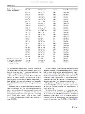
Table 2 GenBank accession Site code Specimen code Taxon GenBank accession numbers
numbers of the studied
specimens
CAL_ST CAL_ST_281 PB2 JX982340
FAV_PO FAV_PO_1 PB1 JX982350
FAV_PO FAV_PO_2 PB1 JX982351
LAM_CF LAM_CF_284 PB1 JX982361
LAM_SC LAM_SC_256 PB1 JX982364
LIN_CP LIN_CP_285 PA JX982354
MAL_GH MAL_GH_11 PB1 JX982360
MAL_GH MAL_GH_6 PB1 JX982344
MAL_RY MAL_RY_283 PA JX982341
MAL_SB MAL_SB_188 PB1 JX982342
MAL_SMB MAL_SMB_308 PB1 JX982343
SIC_CA SIC_CA_8 PA JX982345
SIC_CB SIC_CB_307 PB2 JX982362
SIC_FA SIC_FA_1 PB2 JX982363
SIC_FB SIC_FB_1 PA JX982348
SIC_FB SIC_FB_5 PA JX982349
SIC_MA SIC_MA_305 PA JX982356
SIC_OL SIC_OL_1 PB2 JX982358
SIC_OL SIC_OL_253 PB2 JX982359
SIC_RO SIC_RO_304 PA JX982355
SIC_SM SIC_SM_306 PB2 JX982365
SIC_SV SIC_SV_303 PA JX982353
SIC_TF SIC_TF_282 PB2 JX982347
SIC_TG SIC_TG_2 PA JX982352
SIC_TM SIC_TM_5 PA JX982346
VUL_GE VUL_GE_1 PA JX982357
PA, Phaleria acuminata; PB1,
P. bimaculata ‘‘Southern sub- VUL_GE VUL_GE_309 PA JX982339
clade’’; PB2, P. bimaculata VUL_VP VUL_VP_2 PB2 JX982338
‘‘Tyrrhenian sub-clade’’
i.e., the pronotum and the elytra. Statistical variations in The digital images of the pronotum and the elytra were
the shape of the pronotum and of the right elytra in dif- processed separately with mAKEfAN6(Sheets 2003). For each
ferent P. bimaculata and P. acuminata individuals were body structure, the landmarks and semi-landmarks config-
analyzed using multivariate statistics. uration was identified following criteria of homology
The pattern of morphological body-shape variation was (Bookstein 1991). Within the pronotum, the Cartesian x, y
analyzed in eighteen Phaleria spp. populations. Analyses coordinates of four landmarks and six semi-landmarks were
were performed on 206 pronota and 216 elytra (Table 1). recorded. In the elytra, the Cartesian x, y coordinates of four
Head and limbs from sampled specimens were dissected to landmarks and twelve semi-landmarks were recorded.
be used for the DNA extraction and subsequent molecular The position of the pronotum landmarks and semi-
analyses. landmarks adopted in this study is shown in Fig. 2a, while
After dissection, the remaining body parts were mounted the position of elytra landmarks and semi-landmarks is
on an entomological card. The specimens were positioned shown in Fig. 2b.
along a horizontal plane; for each individual, only the right The bidimensional coordinates of the anatomical land-
portion of the body was examined, with the aim of marks and semi-landmarks on the outline of the dorsal view of
removing any possible bias caused by bilateral asymmetry. the pronotum and the right elytra were collected and digitized
Dorsal images were digitized using a Leica D-LUX by means of TPSDIG2 (Rohlf 2004). In order to better perform
3 LMS camera mounted on the optical stereomicro- statistical analysis, the landmarksand semi-landmarks were
scope Wild M3. successively recognized with TPSUTIL 1.45 (Rohlf 2008).
123